The smallest living entity of an organism is the cell. In contrast to single-celled organisms that are independent entities, the cells of higher organisms form functional units. In accordance with their function, the cells are differentiated by size, shape, and the degree of definition of certain characteristics.
For all cells of the body there are a certain basic structure and numerous basic properties. The basic properties include the ability to divide and to sense and respond to stimuli.
Basic Cell Structure
By and large, the cell consists of the cytoplasm containing the cell organelles, the nucleus, and the cell membrane surrounding the whole structure.
Cell Membrane
The cell membrane, also known as the elementary membrane, consists of a double lipid layer, in which the fat-soluble components face each other while the water-soluble parts form the inner and outer boundaries (a three-layered structure). The lipid molecules are infiltrated with proteins. The outer side of the membrane is covered by a glycocalyx. An elementary membrane also surrounds the cell organelles and the nucleus.
Cytoplasm and Cell Organelles
The cytoplasm consists of the intracellular fluid (cytosol), the cell organelles, and various cell inclusions (phakeroplasm). The cell organelles are responsible for the cell’s metabolism.
Endoplasmic Reticulum (ER)
Present in all cells except erythrocytes; serves intracellular material transport; protein synthesis (granular ER); lipid and hormone synthesis (smooth ER).
Ribosomes
No elementary membrane; multienzyme complexes made up of proteins and rRNA molecules that link amino acid chains for protein synthesis.
Free ribosomes: intracellular proteins (enzymes, etc.). Ribosomes in the ER (granular ER): exported proteins (glandular secretions, etc.).
Golgi Apparatus
Present in all cells except erythrocytes; uptake and discharge products of synthesis in the form of membrane-bounded transport vesicles that are flushed from the cell (secretory vesicles) and serve the renewal of the cell membrane or take part in intracellular digestion as primary lysosomes.
Lysosomes
“Digestive organs” of the cell; with the aid of enzymes they degrade cell-alien structures and the cell’s own decaying organelles.
Centrioles
Build the spindle fibers during cell division.
Mitochondria
“Power stations” of the cell; here nutrients (proteins, fats, carbohydrates) are metabolized essentially to CO2 and H2O, generating the energy necessary for metabolism (e. g., muscle contraction, synthesis of structural substances), which is then stored in the form of ATP.
Cell Nucleus
Present in all cells except erythrocytes; the nucleus contains the nucleolus (production of rRNA ⇒ protein biosynthesis) and the chromosomes, carriers of the hereditary factors (genes). Human nuclei contain 23 chromosome pairs (23 paternal, 23 maternal ⇒ diploid chromosome set); the 23rd pair determines sex.
The appearance of the nucleus and of the chromosomes changes with the individual phases of cell division.
During the interphase (working phase of the cell) between two cell divisions (mitoses) the genetic material is duplicated and chromosomes form, each with two chromatids joined by a constriction (centromere). Each chromatid consists of one molecule of DNA (deoxyribonucleic acid). The basic units contained in DNA, the nucleotides, are each composed of one base (adenine, cytosine, guanine, or thymine), a sugar (deoxyribose), and an acid phosphate radical. DNA contains the complete hereditary matter in the form of genes.
Each unit of information comprises three bases (triplet, codon) in varying combinations. Each triplet represents the information for one amino acid. One gene consists of about 300−3000 base triplets and provides the information for one protein. This genetic code is the same for all living things and contains the information for the biosynthesis of proteins, the most important structural and energizing substances in all organisms.
Protein Biosynthesis
Single-stranded RNA synthesized in the nucleus copies the genetic code (transcription) and brings the message to the ribosomes, the site of protein biosynthesis. Each copied triplet represents one amino acid in the final protein. tRNA molecules, also synthesized in the nucleus, bind amino acids in accordance with the genetic code (according to the sequence of triplets) and transport them to ribosomes, where they are linked into proteins with the aid of enzymes. Each tRNA is specific for one amino acid.
Cell Division (Mitosis)
Chromosomes containing two chromatids are created by the duplication of genetic material during interphase. This process is necessary for the transmission of genetic information to the daughter cells. Mitotic division of cells makes possible growth and the renewal of cells.
Reduction or Maturation Division (Meiosis)
Two successive cell divisions lead to the creation of male or female sex cells with half the chromosome complement (haploid cells).
First maturation division: The (homologous) paternal and maternal chromosomes lying next to each other separate, which leads to the exchange of homologous fragments by “crossing-over.” This results in two daughter cells with haploid chromosome sets.
Second maturation division: Corresponds to a normal mitosis. The chromatids of the chromosomes separate again. The two daughter cells create four mature sex cells with a haploid set of chromosomes.
Fertilization creates a new diploid set of chromosomes. The actual reason for meiosis is the restructuring and recombination of the chromosomes, that is, the shuffling of genetic material.
Intracellular and Extracellular Fluid
The body of an adult person consists of 60% water, two-thirds of which is intracellular, one-third extracellular. Of the 14 liters of extracellular fluid, three-quarters occupy the interstitial spaces and onequarter the vascular system.
Membrane Potential
In the extracellular fluid, sodium is the predominant cation and chloride is the predominant anion; in the intracellular fluid, the predominant cation is potassium and proteins are the predominant anions.
By the differential distribution of ions in the intracellular and extracellular spaces, a potential difference is created across cell membranes (membrane or resting potential). This is caused by the active accumulation of potassium inside the cell (ATP-dependent Na+−K+ pump).
Solid and Fluid Transport
Transport processes between the cells and their environment play an important role in the maintenance of the “internal milieu” (homeostasis). A distinction is made between passive and active (energy-dependent) transport processes. Passive processes include free diffusion (e. g., of O2, CO2, H2O), facilitated diffusion (e. g., of glucose and amino acids in the cells of the intestinal mucosa), osmosis, and filtration (e. g., of glucose and amino acids in the capillaries of the tissue). Active processes include active transport (e. g., of ions) as well as endocytosis and exocytosis (e. g., of proteins).
воскресенье, 31 октября 2010 г.
среда, 27 октября 2010 г.
Endocytosis and Exocytosis
Large molecules, such as proteins, enter (endocytosis) or exit (exocytosis) through the cell membrane by so-called vesicular transport. During this process, substances are attached in part to the outside of the cell by membrane-bound receptors, enclosed by a part of the plasma membrane, and moved into the interior of the cell as a membranewrapped vesicle (receptor-mediated endocytosis). Depending on the size of the absorbed particle, this process may also be called pinocytosis or phagocytosis.
In exocytosis, products synthesized in the cell are enclosed in membranous vesicles and, by coalescence of these vesicles with the inside of the plasma membrane, reach the extracellular space. In this way, the transmitter substances in the endings of nerve cell processes are liberated at the synapses. The secretory products of most glandular cells leave the cell interior in similar fashion. Endocytosis and exocytosis are dependent on the action of ATP.

In exocytosis, products synthesized in the cell are enclosed in membranous vesicles and, by coalescence of these vesicles with the inside of the plasma membrane, reach the extracellular space. In this way, the transmitter substances in the endings of nerve cell processes are liberated at the synapses. The secretory products of most glandular cells leave the cell interior in similar fashion. Endocytosis and exocytosis are dependent on the action of ATP.

Exocytosis and endocytosis
понедельник, 25 октября 2010 г.
Active Transport
Active transport is the transport of substances through the cell membrane by means of an energy-consuming transport system (transport ATPase). Here, again, ATP serves as universal fuel. Such a transport process can move a substance through the membrane against a concentration gradient. Thus cells have the ability to maintain in their interior stable ion concentrations, for example, that are clearly different from their concentrations in the extracellular fluid. These active transport processes are served by specialized proteins in the cell membrane that can move several ions simultaneously. In this process, the coupled transport of substances can occur in the same direction (cotransport) or in opposite directions (countertransport). For instance, in the kidney the transport of amino acids is coupled with an active Na+ transport. Additionally, active ion transport through cell membranes is necessary for the formation of membrane or resting potentials.
воскресенье, 24 октября 2010 г.
Filtration
Filtration occurs when water and any dissolved particles are pushed through cell membranes or pore systems by a hydrostatic pressure difference. Pores occur, for instance, when there are small spaces between endothelial cells (intercellular clefts) or holes (fenestrations) in the cell membranes. Such a process is found in the capillaries of the tissues. The term ultrafiltration is used when, in the course of filtration processes such as that in the capillaries of the renal corpuscles, larger blood components are retained or dissolved molecules are separated out because of their size or charge.
суббота, 23 октября 2010 г.
Osmosis and Osmotic Pressure
When two solutions containing different concentrations of the same solute are separated by a partly permeable membrane, a so-called semipermeable membrane, osmosis can take place. In osmosis the semipermeable membrane allows the solvent, but not the solute, to pass.Water diffuses through the membrane toward the solution of higher concentration, until equilibrium is attained. During this process the volume of the side that initially contained the higher concentration increases. The pressure that must be applied to this side to reverse the process of osmosis is called the osmotic pressure. It is expressed inmmHg or in the SI units of pascals (Pa) or kilopascals (kPa). When such a measurement is applied, it is found that the osmotic pressure depends only on the number of dissolved particles in a defined volume, and not on their size or charge.
The cell membranes are more or less semipermeable membranes, since the lipid layer is less permeable to charged molecules such as ions and proteins. The osmotic pressure of the extracellular fluid depends on its content of protein and salts and corresponds approximately to that of a 0.9% solution of NaCl. Such a physiological salt solution is isotonic (that is, it is in osmotic equilibrium with the cell). Consequently, cells bathed in hypertonic (more concentrated) solutions lose water and shrink, while in hypotonic (less concentrated) solutions they take up water and swell. The organism therefore endeavors by special regulatory mecha nisms to keep the osmotic pressure of the extracellular fluid as constant as possible. Because of the good permeability of cell membranes to water, these mechanisms lead to a more or less constant osmotic pressure in the cell interior.

Thetermcolloidosmoticpressureisusedwhen,for instance, proteins for which the capillary wall is impermeable are dissolved in the blood plasma and not in the interstitial fluid. They create an osmotic pressure difference of about 25mmHg (3.3 kPa) between the interstitial fluid and the capillary space. Thiswould lead to amovement of fluid into the vessels if the hydrostatic blood pressure active inside the blood vessels were not opposed to it. Since the blood pressure at the beginning of the capillaries (37 mmHg)is greaterthanthe colloidosmotic pressure, fluid is actuallyfiltered into the interstitial space.

Development of osmotic pressure in a semipermeable membrane.
вторник, 19 октября 2010 г.
Solid and Fluid Transport
The specific transport processes that take place in the microscopic realm, e. g., between the cells on the one hand and the blood capillaries and their surrounding cells on the other, can be divided into essentially passive (diffusion, osmosis, and filtration) and active (energy-dependent) transport processes (active transport, endocytosis, exocytosis).
понедельник, 18 октября 2010 г.
Membrane or Resting Potential of a Cell
Because the ions are distributed unevenly between the intracellular and extracellular spaces, a potential difference, known as the membrane potential, is created at the cell membrane. This creates a negative charge in the interior of the cell relative to the extracellular space, the so-called resting potential. This potential difference can be measured with sensitive instruments and is about 60−80mV.
The reason for the negative potential inside the cell with respect to its surroundings lies in the differential distribution of ions between the intracellular and extracellular spaces. Thus, the intracellular potassium concentration is about 35 times greater than the extracellular concentration, while proteins are the preponderant anions inside the cell. Sodium ions dominate in the extracellular space, balanced on the negative side by chloride anions. The accumulation of potassium ions inside the cell is a specific activity of almost every cell and represents one of its most important active transport processes. This “ion pump” transports potassium ions into the cells and, to balance this, transports sodium ions out. It is therefore also called the sodium−potassium (Na+−K+) pump. It includes an ATP-splitting enzyme (sodium−potassium ATPase, Na+,K+-ATPase). This reaction liberates the energy required for ion transport. The cell membrane is impermeable to ions, so there are membrane pores (channels) forNa+, K+, and Cl−, but not for protein anions. During resting potential, the K+ channels are often open, but the Na+ and Cl− channels are mostly closed. Because of the concentration difference, the K+ ions have a tendency to diffuse outward. However, the diffusion of positively charged potassium ions out of the cell is limited by the negatively charged protein anions, which cannot cross the membrane because of their size. The diffusion of even a few potassium ions out of the cell leaves anions with the opposite (negative) charge (protein anions) on the inside of the cell membrane, so that the interior of the cell is negatively chargedwith respect to its surroundings. The resting potential is therefore also known as the diffusion potential. The diffusion of ions outwardthrough the membrane pores is independent of the Na+−K+ pump.
The energy-consuming ion pumps can be impeded or blocked by lack of oxygen (failure of ATP production) or by metabolic poisons (e. g., cyanide), leading to severe disturbances in the specific performance of a cell. The initiation and propagation of nerve or muscle cell excitation depends on brief membrane potential changes (action potentials) (see Chapter 3: Nerve Tissue).

суббота, 16 октября 2010 г.
Composition of the Intracellular Fluid
In contrast to the extracellular fluid, where sodium predominates, the quantitatively dominant intracellular cation is potassium (K+). The sodium concentration inside the cell is about 10 times smaller than that outside it. The major portion of the intracellular anions consists of proteins, while phosphates (HPO4
−/H2PO4 −) are present in lower concentrations.
−/H2PO4 −) are present in lower concentrations.
пятница, 15 октября 2010 г.
Composition of the Extracellular Fluid
The substances dissolved in the extracellular fluid (e. g., salts) are present as electrically charged particles (ions) and the solutions are called electrolytes. Because of their electric charge, ions can migrate in electric fields. For this reason, positively charged ions are also called cations (they migrate to the negative pole, the cathode) and negatively charged ions are called anions (they migrate to the positive pole, the anode). The salt present in the largest amount is common table salt (NaCl), consisting of a positively charged sodium ion (Na+) and a negatively charged chloride ion (Cl−) dissolved in a concentration of about 9 g/l. Other cations and anions are also present, though in distinctly smaller quantities: e. g., potassium (K+), calcium (Ca2+) and magnesium (Mg2 +), as well as
bicarbonate (HCO3 −) and negatively charged proteins. The three compartments of the extracellular space—the interstitial fluid, the blood plasma, and the lymph—differ mainly in the amount of protein dissolved in each. For instance, the walls of the blood and lymph capillaries are permeable only to small ions and smaller organic particles, while large proteins are retained within the lumina of these vessels.
bicarbonate (HCO3 −) and negatively charged proteins. The three compartments of the extracellular space—the interstitial fluid, the blood plasma, and the lymph—differ mainly in the amount of protein dissolved in each. For instance, the walls of the blood and lymph capillaries are permeable only to small ions and smaller organic particles, while large proteins are retained within the lumina of these vessels.
среда, 13 октября 2010 г.
Exchange of Materials between the Cell and Its Environment
Billions of years ago, life developed in the form of small, single-celled organisms in a large primal ocean. Their aqueous (watery) environment was marked by a milieu of constant composition. Nutrients were plentiful, and waste products were instantly diluted effectively to infinity. In a similar way, the cells of a multicellular organism live in an aqueous environment that contains all the salts and nutrients required for the sustenance of the cell. Compared to the primal ocean, however, this fluid has a much smaller volume, and there is a much greater danger of short-term changes in its composition.
Of all the chemical compounds in the organism, water (H2O) forms the largest percentage part. Thus the body of an adult contains about 60% water, which is distributed in two distinct compartments: the intracellular space (total volume enclosed in all the cells) and the extracellular space (total volume present outside the cells). About two-thirds of the total body fluids are located inside the cells (intracellular fluid) and the remaining third (about 14 liters in a person weighing 70 kg) bathes the exterior of the cells. Of the 14 liters of extracellular (interstitial) fluid, three-quarter are contained in the tiny spaces that separate the cells from each other, and one-quarter in the vascular systems (arteries, veins, capillaries, and lymphatic vessels), where it forms the aqueous part of the plasma and the lymphatic fluid.
 The environment (milieu) in which the cell lives. (After Silbernagel)
a Unicellular organism: Interaction between the first cells and their environment—the primal ocean, a milieu distinguished by its constant composition
b Human: Cells in a multicellular organism are bathed in extracellular fluid, the volume of which is distinctly smaller than that of the fluid inside the cell. This “internal milieu” would change its composition very quickly if the space between the cells (interstitial space, intercellular space) were not connected to organs such as the lung, the kidney, or the digestive tract by the vascular bed, which take up fresh nutrients and eliminate metabolic waste products.
The water content of the body is kept constant with great precision. This is necessary in order not to endanger the equilibrium of the numerous substances dissolved in the body fluids. For instance, physiological water losses (e. g., the production of urine, the secretion of sweat, and loss by humidification of expired air) must be balanced by fluid intake.
Keeping the “internal milieu” constant (homeostasis) is a life-preserving precondition for the optimal functioning of every cell in the body. Since the most diverse substances reach the extracellular space as a result of respiration, the intake of nutrients, and the metabolic activity of cells, maintaining homeostasis is one of the most important tasks of the organism. Besides the activity of the lungs, the intestines, and the kidneys, certain specific transport processes are of importance; these processes (e. g., diffusion, osmosis, active transport) serve to exchange solids and fluids between the cell and its environment (see Solid and Fluid Transport below). Transport of substances over greater distances within the body (e. g., nutrients taken up in the intestines and oxygen taken up in the lungs) is accomplished in the blood vessels. Similarly, transport by the lymphatics, passage through the intestines, and the emptying of the gallbladder accomplish rapid distribution of solids and fluids.
Of all the chemical compounds in the organism, water (H2O) forms the largest percentage part. Thus the body of an adult contains about 60% water, which is distributed in two distinct compartments: the intracellular space (total volume enclosed in all the cells) and the extracellular space (total volume present outside the cells). About two-thirds of the total body fluids are located inside the cells (intracellular fluid) and the remaining third (about 14 liters in a person weighing 70 kg) bathes the exterior of the cells. Of the 14 liters of extracellular (interstitial) fluid, three-quarter are contained in the tiny spaces that separate the cells from each other, and one-quarter in the vascular systems (arteries, veins, capillaries, and lymphatic vessels), where it forms the aqueous part of the plasma and the lymphatic fluid.

a Unicellular organism: Interaction between the first cells and their environment—the primal ocean, a milieu distinguished by its constant composition
b Human: Cells in a multicellular organism are bathed in extracellular fluid, the volume of which is distinctly smaller than that of the fluid inside the cell. This “internal milieu” would change its composition very quickly if the space between the cells (interstitial space, intercellular space) were not connected to organs such as the lung, the kidney, or the digestive tract by the vascular bed, which take up fresh nutrients and eliminate metabolic waste products.
The water content of the body is kept constant with great precision. This is necessary in order not to endanger the equilibrium of the numerous substances dissolved in the body fluids. For instance, physiological water losses (e. g., the production of urine, the secretion of sweat, and loss by humidification of expired air) must be balanced by fluid intake.
Keeping the “internal milieu” constant (homeostasis) is a life-preserving precondition for the optimal functioning of every cell in the body. Since the most diverse substances reach the extracellular space as a result of respiration, the intake of nutrients, and the metabolic activity of cells, maintaining homeostasis is one of the most important tasks of the organism. Besides the activity of the lungs, the intestines, and the kidneys, certain specific transport processes are of importance; these processes (e. g., diffusion, osmosis, active transport) serve to exchange solids and fluids between the cell and its environment (see Solid and Fluid Transport below). Transport of substances over greater distances within the body (e. g., nutrients taken up in the intestines and oxygen taken up in the lungs) is accomplished in the blood vessels. Similarly, transport by the lymphatics, passage through the intestines, and the emptying of the gallbladder accomplish rapid distribution of solids and fluids.
воскресенье, 10 октября 2010 г.
Schematic representation of maturation division (meiosis).
In order to provide a better overview, the course of the two maturation divisions is exemplified in a germ cell with three pairs of chromosomes (red chromosomes = paternal, blue chromosomes = maternal). During the pachytene of the prophase of the first maturation division, the chromatids become visible. The maternal and paternal chromosomes arrange themselves next to each other and form a tetrad (two chromosomes each with two chromatids). During this process the paternal and maternal chromatids partly overlap and when they separate there is an interchange of fragments (“crossing-over”). During themetaphase of the first maturation division, the homologous (paternal and maternal) chromosomes separate, and are randomly distributed to the two daughter cells. This process creates two haploid daughter cells each with a single set of chromosomes. During the second maturation division, the two daughter chromatids separate by mitotic cell division, so that the first and second maturation divisions result in the creation of four haploid sex cells. (After Beske)
пятница, 8 октября 2010 г.
Reduction or Maturation Division (Meiosis)
Reduction or maturation division (meiosis) is a special form of cell division. In preparation for later fertilization, the male and female germ cells must halve their set of chromosomes (to form a haploid set), so that when ovum and sperm join, a normal double (diploid) set of chromosomes is formed. This process is known as meiosis, a cell division that comprises two steps: first and second maturation divisions.
Shortly before the first maturation division, the male and female sex cells duplicate their DNA as in mitosis, with each chromosome containing two identical chromatids. In meiosis, the prophase of the first maturation division lasts a good deal longer than the prophase of mitosis. As a rule it takes 24 days in male germ cells, while in the female it may at times take decades, because of an interpolated resting phase (dictyotene) (see Chapter 11: Development of the Ovum (Oogenesis) and Follicle Maturation). Prophase is divided into five phases: leptotene, zygotene, pachytene, diplotene, and diakinesis.


First maturation division. During the leptotene of the prophase, the chromosomes become visible as fine threads; in the ensuing zygotene, they arrange themselves side by side in pairs (chromosome pairing). During this process, the corresponding (homologous) paternal and maternal chromosomes are always arranged next to each other. Since each single chromosome contains two chromatids (sister chromatids), the chromosome pairs contain four chromatids, two maternal and two paternal, the so-called tetrad, which is especially conspicuous during the diplotene of prophase. At this point the homologous chromosomes begin to separate. During this process, homologous paternal and maternal chromatids lying in parallel next to each other can interchange homologous fragments by so-called crossing-over (forming a chiasma) and reattachment of the fragments.
In the metaphase which follows, the chromosomes arrange themselves in the equatorial plane, similarly to a mitosis. During anaphase, the separation of the homologous chromosomes by a spindle is completed. Telophase completes the first maturation division. The two daughter cells each now have only half the number of chromosomes of the initial cell, though each chromosome still consists of two chromatids.
Second maturation division. After a brief phase (interkinesis) during which the DNA is not duplicated, the second maturation division begins. This maturation division proceeds entirely like a normal mitosis, that is, during anaphase the two chromatids of each chromosome divide and are distributed to two daughter cells. Consequently, the haploid daughter cells that originated when the double set of chromosomes was halved during the first maturation division, once again halve their DNA content during the second maturation division.
The result of the two maturation divisions = mature sex cells. Thus four daughter cells (mature sex cells) result from the two-step division of meiosis. In each of these, the number of chromosomes and the DNA content have been reduced to half the original. In addition, the chromosomes have been restructured as a result of the crossing-over, and recombined as a result of the random distribution of the two homologous chromosomes into the daughter cells. The real biological significance of meiosis lies in this shuffling of genetic material.
вторник, 5 октября 2010 г.
Course of a Mitosis
At the beginning of prophase the cell becomes round and the chromosomes appear in the nucleus as convoluted threadlike structures. Simultaneously, the nuclear membrane disappears and the two centrioles move apart. They migrate toward the poles of the cell and form the socalled central spindle. In metaphase, which follows, the chromosomes become shorter and thicker, the two chromatids become visible and can be distinguished clearly by their size and shape. As the process continues, the chromosomes arrange themselves in the so-called equatorial plane between the two poles.

Schematic representation of cell division (mitosis) (After Hadorn and Wehner)
a Prophase: Chromosomes in the nucleus become visible by coiling and the nuclear spindle apparatus develops the central spindle;
b Early metaphase: the central spindle stretches and the chromosomes migrate toward the equatorial plane;
c Late metaphase: The division of each chromosome into two chromatids is clearly visible and arrangement along the spindle’s equator is complete;
d, e Anaphase: the daughter chromosomes are moving away from each other in the direction of the spindle’s pole;
f Telophase: the chromosomes have uncoiled, a nuclear membrane has formed and the cell body is constricted.
At the end of metaphase the chromosomes have arranged themselves in the equatorial plane in such a way that each of their constrictions (centromere) is oriented toward the central axis. Because the arrangement looks star-shaped when viewed from the two poles, it is called a “monaster” (Greek for single star). As anaphase begins, the chromatids (chromosome halves) of each chromosome separate, forming two starshaped figures called “diasters” (double star). Since each of the halves of a chromosome (daughter chromatids) migrates to one or other of the two opposite poles, all of the genetic material is divided equally between the two daughter cells.
In the ensuing telophase the chromatids, which now form the chromosomes of the two daughter cells, collect near the centrioles, uncoil, and again become invisible. As a newnuclear membrane forms, two new interphase nuclei have been created. There follows a complete division of the cell body, resulting essentially in two equal, independent daughter cells.
On average one mitosis takes about 60 minutes to complete. Anaphase is the shortest phase, lasting about 3 minutes.
суббота, 2 октября 2010 г.
Cell Division (Mitosis)
Duplication (replication) of DNA and the transmission of genetic information to the two daughter cells connected with it precede every cell division and take place during the so-called interphase. The interphase is the stage between mitoses and is the working phase of the cell. Chromosomes with two chromatids are formed by duplication of the genetic material during interphase. This establishes the precondition for mitotic cell division. Chromosomes demonstrate evidence of their duplication by a longitudinal split visible in the microscopic image. The chromosomes become shorter and thicker through increased coiling. After cell division is complete, the chromosomes uncoil and, during the ensuing interphase, replicate again.
Mitotic cell divisions permit a fertilized ovum to develop into an organism. They are the preconditions for physiological renewal of cells and lead to regeneration of tissues after injury. With the exception of a fewcells (nerve cells, and cardiac and skeletal muscle cells), the ability to divide is maintained throughout the life cycle, though it varies among cells. As a rule, mitoses are less common in highly differentiated tissues.
Mitotic cell divisions permit a fertilized ovum to develop into an organism. They are the preconditions for physiological renewal of cells and lead to regeneration of tissues after injury. With the exception of a fewcells (nerve cells, and cardiac and skeletal muscle cells), the ability to divide is maintained throughout the life cycle, though it varies among cells. As a rule, mitoses are less common in highly differentiated tissues.

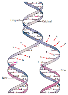
Double helix of DNA and its duplication (replication). The double strand of DNA splits like a zipper and forms two new, completely identical DNA molecules. The original strands are shown in blue, the new ones in pink (the sugar−acid phosphate chain is shown as a band; A = adenine, C = cytosine, G = guanine, T = thymine; free nucleotide groups are marked by arrows) (After Hadorn and Wehner)
Подписаться на:
Сообщения (Atom)