Basic Structure
Examination of a cell by light microscopy shows a fluid cell body (cytoplasm), a cell nucleus and the surrounding cell membrane (plasmalemma). The cytoplasm contains a number of highly organized small bodies, called cell organelles, that can often only be seen by electron microscopy. It also contains certain supportive structures (parts of the cytoskeleton) and numerous cell inclusions (e. g., metabolic substrates and end products).
Cell Membrane
The surrounding cell membrane (plasmalemma) contains the fluid cell body (protoplasm). An electron-microscopic section demonstrates a three-layered structure: this includes a double layer of lipids in which two layers of lipid molecules (phospholipids, cholesterol), are arranged so that their lipid-soluble parts (fatty acids) oppose each other (light middle line) while the water-soluble ends form the outer and inner boundaries of the cell membrane (dark outer and inner lines). The double lipid layer is infiltrated with proteins in a more or less mosaiclike fashion. These protein molecules have multiple functions. They may form pores that serve the transmission of water and salts, or they may take part in regulatory functions as receptor proteins. The membrane proteins abutting on the outer side of the cell, and in part the watersoluble ends of the phospholipids, are covered with a thin film of sugar molecules (carbohydrates). This film is called the glycocalyx. The chemical structure of the glycocalyx is laid down genetically and it is specific for each cell. By this structure cells can “recognize” each other as self and non-self (see Chapter 6: The Immune System, Specific Immunity). This so-called elementary membrane has a thickness of 7.5 nm (1 μm = 1000 nm) and forms a barrier between the cell interior and the extracellular space. The cell organelles are also surrounded by elementary membranes.

Simplified image of a cell representing electron-microscopic findings
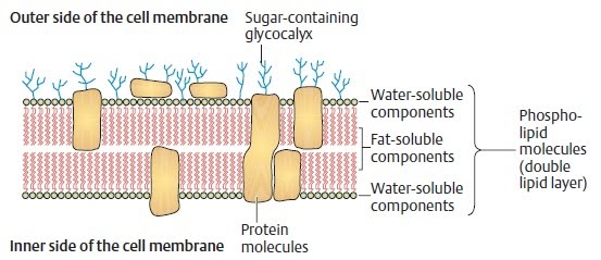
Schematic cross-section of a cell membrane. The three-layered structure seen
in the electron-microscopic image is produced by the two water-soluble inner and outer
components of the double lipid layer, and the fat soluble components between them.
(After Leonhardt)
Cytoplasm and Cell Organelles
The cytoplasm surrounds the cell nucleus. It is composed of the hyaloplasm or cytosol (intracellular fluid), the cell organelles that perform certain cellular functions, and various cell inclusions, the metaplasm (metabolic products of the cell). The intracellular fluid consists of an aqueous saline solution and proteins (microtubules, microfilaments, intermediate filaments) that determine the shape and mechanical solidity of the cell (the so-called cytoskeleton). The organelles vary in number according to the type and function of the cell containing them. The following essential cell organelles may be differentiated:
Endoplasmic reticulum
Ribosomes
Golgi apparatus
Lysosomes
Centrioles
Mitochondria
Endoplasmic Reticulum (ER)
The endoplasmic reticulum criss-crosses the cytoplasm in the form of tubular and vesicular structures surrounded by elementary membranes. It subdivides the interior of the cell into compartments and facilitates the intracellular transport of substances along its channels. Its large surface makes possible the rapid completion of specific metabolic processes (e. g., the synthesis of proteins and lipids) and serves as a depot for membranes, i.e., it originates other membranes. In many places the endoplasmic reticulum is dotted with small granular structures, the ribosomes (granular ER), that serve especially for the synthesis of proteins (see below). Granular endoplasmic reticulum is especially prominent in cells such as those of the pancreas. The endoplasmic reticulum is called smooth ER when ribosomes are absent, predominating especially in hormone-secreting cells. All cells except red blood cells contain endoplasmic reticulum.
Ribosomes
Ribosomes serve protein synthesis (see also The Cell Nucleus, Protein Synthesis below) and occur either separately in the form of free ribosomes or in combination with endoplasmic reticulum (granular ER). They are not surrounded by elementary membrane. In the granular ER they are responsible for the production of exported proteins (e. g., glandular secretions), whereas free ribosomes produce intracellular proteins (e. g., enzymes, structural proteins). Ribosomes contain complexes made up of several enzymes consisting of proteins and RNA molecules (ribosomal RNA, rRNA). These create the amino acid chains for protein synthesis. rRNA is also a structural element of ribosomes.
Комментариев нет:
Отправить комментарий